45 ear anatomy without labels
Image result for ear structure without label 2018. 2. 12 - Image result for ear structure without label. 2018. 2. 12 - Image result for ear structure without label. Pinterest. Today. Explore. ... See 12 Best Images of Anatomy Human Ear Diagram Worksheet. Inspiring Anatomy Human Ear Diagram Worksheet worksheet images. Holly Crimm. School. Middle School Science. Ear Anatomy without Labels, Digital Art - Shutterstock Ear Anatomy Without Labels Digital Art Stock Illustration 530108302 Edit Download for free See more Popularity score High Usage score High usage Superstar Shutterstock customers love this asset! Item ID: 530108302 Ear Anatomy without Labels, Digital Art Formats 8976 × 6201 pixels • 29.9 × 20.7 in • DPI 300 • JPG
Applied Sciences | Free Full-Text | A Novel Immersive Anatomy ... Jun 03, 2022 · Immersive technologies are redefining ways of interacting with 3D objects and their environments. Moreover, efforts in blended learning have presented several advantages of incorporating educational technology into the learning space. The advances in educational technology have in turn helped to widen the choice of different pedagogies for improving learner engagement and levels of ...

Ear anatomy without labels
Radiological anatomy of the spine - e-Anatomy - IMAIOS Sep 13, 2021 · This radioanatomy module of the spinal column presents 18 conventional radiographs of the spine with 192 anatomical structures labeled. It is particularly useful for radiologists, electroradiology students, emergency physicians, orthopedic surgeons and rheumatologists, but may be used as a daily or a teaching support for any practitioner, physician or student involved in the musculoskeletal ... Ears: Facts, Function & Disease | Live Science The external ear, also called the auricle or pinna, is the loop of cartilage and skin that is attached to the outside of the head. It works much like a megaphone. Sound waves are funneled through... Ear Anatomy: Understanding the Outer, Middle, and Inner Parts of the Ear The external auditory meatus, or ear canal, is a narrow canal that leads from the concha to the tympanic membrane, or eardrum. Sound waves are delivered through this canal. This canal is prone to ear infections. Tragus This is the small, rigid part of the ears along the front of the ear, adjacent to the face.
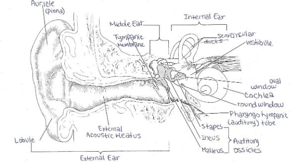
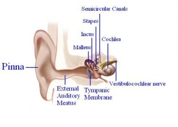
Ear anatomy without labels. Picture of the Ear: Ear Conditions and Treatments - WebMD The ear has external, middle, and inner portions. The outer ear is called the pinna and is made of ridged cartilage covered by skin. Sound funnels through the pinna into the external auditory... Human Ear Anatomy - Parts of Ear Structure, Diagram and Ear Problems The external (outer) ear consists of the auricle, external auditory canal, and eardrum (Figure 1 and 2). The auricle or pinna is a flap of elastic cartilage shaped like the flared end of a trumpet and covered by skin. The rim of the auricle is the helix; the inferior portion is the lobule. Ligaments and muscles attach the auricle to the head. Ear Anatomy Diagram - EnchantedLearning.com hammer - (also called the malleus) a tiny bone that passes vibrations from the eardrum to the anvil. nerves - these carry electro-chemical signals from the inner ear (the cochlea) to the brain. outer ear canal - the tube through which sound travels to the eardrum. pinna - (also called the auricle) the visible part of the outer ear. Ear Anatomy, Diagram & Pictures | Body Maps - Healthline Outer ear: The outer ear includes an ear canal that is is lined with hairs and glands that secrete wax. This part of the ear provides protection and channels sound. The auricle or pinna is the most...
Cefdinir Antibiotic Side Effects, Uses (Strep, Middle Ear ... Cefdinir is an antibiotic in the cephalosporin drug class prescribed to treat infections, for example, middle ear, tonsillitis, strep throat, bronchitis, and sinusitis. Common side effects are nausea, abdominal pain, loose stools, and vaginitis. Dosage and pregnancy and breastfeeding safety information are included. (PDF) LIBRO PARA COLOREAR NETTER - Academia.edu Enter the email address you signed up with and we'll email you a reset link. "Outer Ear Anatomy Colorful Pattern Without Center Label" Shop "outer ear anatomy colorful pattern without center label" search results for the very best in custom shoes, sneakers, apparel, and accessories by independent artists. The Ear: Anatomy, Function, and Treatment - Verywell Health Essential organs of human hearing and balance, the ears are located on either side of the head, at the level of the nose. Separated into an inner, middle, and outer ear, each ear is an intricate and complicated mixture of bones, nerves, and muscles.
Image result for ear structure without label | Ear diagram, Human ear ... Feb 12, 2018 - Image result for ear structure without label. Feb 12, 2018 - Image result for ear structure without label. Pinterest. Today. Explore. When autocomplete results are available use up and down arrows to review and enter to select. Touch device users, explore by touch or with swipe gestures. ... Anatomy Coloring Pages Pittsburgh ... Ear Anatomy - Outer Ear | McGovern Medical School The medical term for the outer ear is the auricle or pinna. The outer ear is made up of cartilage and skin. There are three different parts to the outer ear; the tragus, helix and the lobule. EAR CANAL The ear canal starts at the outer ear and ends at the ear drum. The canal is approximately an inch in length. Parts of the Ear Labelled Diagram Activity - Twinkl The first worksheet presents an ear with annotations showing the first letters of its key features. For example, a label marked 'P' links to the Pinna (outer ear). The second page shows an ear diagram without labels. The final page shows the labels linking to the beginning letters of each feature, but without the words list. Human Ear Diagram - Bodytomy Auditory Ossicles: The three small bones in the middle ear, called malleus, stapes, and incus, are connected. These bones together are called the auditory ossicles, and their purpose is to let the sound that strikes the eardrum, further into the inner ear.

The Confusing Evolution of Bat Echolocation – Koryos Writes
Ear Diagram Quiz - ProProfs Try this amazing Ear Diagram Quiz quiz which has been attempted 5940 times by avid quiz takers. Also explore over 13 similar quizzes in this category. ... Have you been having pain from an ear infection that will come on fast without improving for several days? This may be due to a couple of reasons and one way to self-diagnose that pain is by ...

Trigger Points Set Exam-Room Anatomy Posters – ClinicalPosters
Ear (Anatomy): Overview, Parts and Functions | Biology Dictionary The human ear picks up and interprets high-frequency vibrations of air, while the sound-sensing organs of aquatic animals are designed to pick up high-frequency vibrations in water. Most vertebrates have two ears: one on either side of the head. In some animals, including most mammals, the ear is also used for balance.
Print Exercise 25: Special Senses - Hearing and Equilibrium flashcards | Easy Notecards
Outer Ear: Anatomy, Location, and Function - Verywell Health Fossa, superior crus, inferior crus, and antihelix: These sections make up the middle ridges and depressions of the outer ear. The superior crus is the first ridge that emerges moving in from the helix. The inferior crus is an extension of the superior crus, branching off toward the head. The antihelix is the lowest extension of this ridge.

Pin by Victoria Pigeon on Free time | Human skull anatomy, Skull anatomy, Skull
Human Ear: Structure and Anatomy - Online Biology Notes Ear ossicles: The three ear ossicles (malleus, incus and stapes) form a chain of lever extending from tympanic membrane to inner ear. The ear ossicles transmit sound wave from ear drum to inner ear. Ear ossicles communicate the ear drum with internal ear through fenestra ovalis ( oval window). The ear ossicles are;
2012 Group Project 6 - Embryology
File:Ear-anatomy-text-small-en.svg - Wikimedia Commons Captions. Description. Ear-anatomy-text-small-en.svg. English: human ear anatomy with detailed diagram. Date. 29 June 2003 (original upload date) Source. Made by Iain 05:39 29 Jun 2003 (UTC) Transferred from en.wikipedia to Commons by Papa November using CommonsHelper.
Post a Comment for "45 ear anatomy without labels"